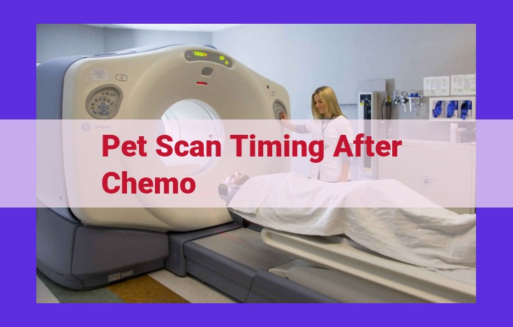
Optimal timing for PET scans after chemo varies based on guidelines and patient factors. Chemo affects glucose metabolism, influencing scan results. Standardized scan timing ensures accurate diagnosis. Inter- and intra-patient variability in timing can impact accuracy. PET scans monitor treatment response, predict outcomes, and assess metabolic changes. Repeated scans track disease progression and metabolic alterations. Chemo-induced metabolic changes and specific chemo agents influence timing protocols.
Optimal Timing of PET Scans After Chemotherapy: A Guide for Enhanced Accuracy
In the realm of cancer diagnosis and monitoring, Positron Emission Tomography (PET)/Computed Tomography (CT) scans play a pivotal role. Understanding the appropriate timing of PET scans after chemotherapy can significantly enhance the accuracy and effectiveness of these scans in detecting and assessing treatment response.
Optimal Time Frame for PET Scans
National and international guidelines recommend performing PET/CT scans 2-4 weeks following the completion of chemotherapy. This time frame allows for post-chemotherapy recovery and restoration of glucose metabolism, ensuring that the scan accurately reflects the metabolic activity of the cancerous cells.
Factors Influencing Scan Timing
The optimal timing of PET scans can vary depending on several factors, including:
- Patient-Related Factors: Blood glucose levels, comorbidities, and nutritional status can impact glucose metabolism and thus scan results.
- Chemotherapy Characteristics: The type and dosage of chemotherapy agents used can affect glucose uptake and metabolism.
- Imaging Protocol: Standardized scan protocols, including fasting guidelines and time after contrast administration, are essential for consistency and accuracy.
Effects of Chemotherapy on Glucose Metabolism
Chemotherapy can significantly alter glucose metabolism within cancerous cells. This can lead to reduced glucose uptake and altered PET scan results, potentially affecting diagnostic accuracy. Understanding these effects is crucial for accurate scan interpretation.
Importance of Standardized Timing
Standardizing scan timing is essential to ensure consistency, reduce variability, and enhance the accuracy of PET scans. This includes adhering to established guidelines for timing, post-chemotherapy recovery intervals, and post-meal intervals.
Impact on Diagnostic Accuracy
Variations in scan timing can impact the sensitivity and specificity of PET/CT scans. Early scans may lead to false negatives due to residual chemotherapy-induced metabolic changes, while late scans may result in false positives due to slow metabolic recovery.
Inter- and Intra-Patient Variability
Variability in scan timing can occur within and between patients. This variability can arise from differences in patient characteristics, chemotherapy regimens, and imaging protocols. Understanding these sources of variability is important for accurate interpretation of PET scans.
Correlation with Treatment Response
PET scans can be valuable in monitoring treatment response to chemotherapy. By comparing scans before and after treatment, clinicians can assess metabolic changes, predict outcomes, and identify areas of treatment resistance.
Factors Affecting PET Scan Timing After Chemotherapy
Patient-Related Factors
The timing of PET scans after chemotherapy is influenced by various patient-specific factors. The patient’s glucose levels play a crucial role as chemo can alter glucose metabolism. Elevated glucose levels can interfere with PET scan accuracy as glucose uptake is used to assess metabolic activity. Therefore, patients are often advised to fast before the scan to ensure optimal glucose levels.
Additionally, patient comorbidities can impact scan timing. For instance, patients with diabetes, impaired glucose tolerance, or renal insufficiency may require specific protocols to account for their underlying conditions. Understanding these factors helps tailor the timing of PET scans to improve diagnostic accuracy.
Chemotherapy Characteristics
The type and stage of chemotherapy administered also influence scan timing. Different chemo agents have varying effects on glucose metabolism. Some agents, such as fluorouracil, can cause rapid metabolic changes, necessitating earlier scans. In contrast, agents like gemcitabine have a more prolonged impact, requiring later scans to capture their effects.
Moreover, the chemotherapy regimen can affect scan timing. Patients receiving neoadjuvant chemotherapy (before surgery) may require shorter intervals between chemo and PET scans to assess tumor response. Conversely, patients undergoing adjuvant chemotherapy (after surgery) may have longer intervals to allow for post-surgical recovery.
Imaging Protocol Elements
The imaging protocol itself can influence scan timing. The type of imaging tracer used (e.g., FDG, F-choline) affects the optimal timing after chemotherapy. Tracer-specific characteristics, such as uptake kinetics and tissue distribution, guide the timing to obtain the most accurate results.
Standardization of Timing
To ensure consistency and comparability of PET scan results, standardization of scan timing is essential. Guidelines recommend specific time frames after chemotherapy, considering patient-specific factors, chemo characteristics, and imaging protocol elements. Adhering to standardized protocols reduces variability and improves the reliability of PET scans for monitoring treatment response and assessing disease status.
Chemotherapy’s Metabolic Impact: How it Alters PET Scan Results
Chemotherapy, a mainstay of cancer treatment, not only targets cancerous cells but also has a significant impact on the body’s metabolism. One key effect is its influence on glucose metabolism, a process crucial for PET/CT scans that aid in cancer diagnosis.
During a PET/CT scan, radioactive glucose is injected into the body. Cancer cells, with their increased metabolic activity, avidly take up this radioactive glucose, emitting signals detected by the scanner. These signals provide valuable information about the tumor’s presence, size, and metabolic status.
However, chemotherapy can disrupt this metabolic process, leading to false negatives or false positives in PET/CT scans. Chemotherapy drugs often deplete glucose stores in the body, essentially “starving” the cancer cells. This metabolic disruption can hinder the uptake of radioactive glucose, resulting in an underestimation of tumor activity. Conversely, chemotherapy-induced inflammation can stimulate glucose uptake in non-cancerous tissues, leading to false-positive readings.
Understanding these metabolic effects is critical for accurate interpretation of PET/CT scans after chemotherapy. Optimizing scan timing, considering chemotherapy-induced metabolic changes, and interpreting results in light of individual patient factors are necessary for reliable and effective cancer management.
Standardizing PET Scan Timing after Chemotherapy
Navigating the complexities of cancer treatment requires precision and personalization. The timing of PET/CT scans after chemotherapy is a crucial factor that impacts the accuracy and effectiveness of diagnosis. Optimizing scan timing involves adhering to standardized guidelines and considering patient-specific characteristics.
Establishing Guidelines
Consensus guidelines provide a framework for standardizing scan timing. _The optimal time frame for PET/CT scans is typically 8-12 weeks after completing chemotherapy_. This window allows adequate time for metabolic recovery and normalization, minimizing the potential for false positives due to treatment-induced metabolic alterations.
Post-Chemo Recovery Intervals
The post-chemotherapy recovery period is critical for restoring normal metabolic function and glucose uptake patterns. _**Scans performed too soon after chemo may underestimate tumor activity due to suppressed glucose metabolism_. Allowing a sufficient recovery interval ensures that metabolic processes have stabilized, providing a more accurate representation of tumor status.
Post-Meal Intervals
Another important factor in scan timing is the _post-meal interval_. Ingesting food can stimulate glucose uptake, potentially interfering with the interpretation of PET/CT scans. _**Patients should typically fast for 4-6 hours before the scan to minimize background glucose activity_. This fasting period ensures that the tracer is primarily taken up by tumors, enhancing the visibility of metabolic abnormalities.
Benefits of Standardization
Standardizing PET scan timing offers several benefits. It:
- Ensures comparability of results over time and between patients
- Reduces the likelihood of false positives and negatives
- Improves diagnostic accuracy and confidence
- Facilitates the monitoring of treatment response and disease progression
- Optimizes patient care by enabling personalized treatment decisions
By following standardized guidelines for PET scan timing, clinicians can enhance the accuracy and value of this vital diagnostic tool, ultimately contributing to better patient outcomes.
Impact of Scan Timing on Diagnostic Accuracy
The timing of PET scans after chemotherapy is crucial for ensuring accurate diagnostic results. Variations in scan timing can significantly affect the sensitivity and specificity of PET/CT scans, potentially leading to false positives and false negatives.
False Positives: When PET scans are performed too soon after chemotherapy, the altered glucose metabolism caused by the treatment can lead to increased FDG uptake, even in non-cancerous tissues. This can result in false positives, where healthy tissues appear cancerous.
False Negatives: Conversely, if PET scans are performed too late after chemotherapy, the glucose metabolism in cancerous tissues may have normalized, leading to decreased FDG uptake. This can result in false negatives, where cancerous tissues are missed.
The optimal timing of PET scans helps to minimize both false positives and negatives, maximizing the accuracy of the results. Standardized guidelines and patient-specific factors should be considered to determine the most appropriate time frame for each individual.
Inter- and Intra-Patient Variability in Scan Timing Impacts Accuracy
PET scans, often performed after chemotherapy, play a crucial role in cancer diagnosis and treatment monitoring. However, scan timing heavily influences the interpretation of results, and variations can occur between and within patients, affecting diagnostic accuracy.
Inter-patient variability arises due to individual differences in glucose metabolism, comorbidities, and medication regimens. The rate at which glucose is taken up and metabolized by cancer cells varies, impacting the timing of optimal scan sensitivity. Comorbidities, such as diabetes, can also affect glucose levels, influencing scan interpretation.
Intra-patient variability refers to variations in scan timing within the same patient. Factors like food intake, exercise, and emotional stress can alter glucose metabolism, leading to differences in PET scan results. Post-chemotherapy recovery periods can also impact scan timing, as metabolic alterations persist after treatment.
Ignoring variability can result in false positives or negatives. For example, if a scan is performed too soon after chemotherapy, remaining glucose uptake in recovering cancer cells may mimic tumor activity. Conversely, delayed scans may miss metabolically active tumors due to decreased glucose uptake.
Standardization of scan timing, including post-chemotherapy recovery intervals and post-meal intervals, is essential to minimize variability and improve accuracy. By optimizing scan timing based on patient-specific factors and standardized protocols, healthcare professionals can enhance diagnostic certainty and ensure reliable assessment of treatment response.
Correlation with Treatment Response
PET scans have proven invaluable in monitoring treatment response to chemotherapy. They offer a unique window into metabolic changes within tumors, allowing clinicians to assess the effectiveness of treatment and predict outcomes.
PET scans can detect metabolic alterations in tumors as early as 24 hours after chemotherapy administration. By measuring the uptake of glucose, a vital energy source for cancer cells, PET scans can determine whether the treatment is inhibiting tumor metabolism. A significant reduction in glucose uptake following chemotherapy is often indicative of tumor shrinkage and a favorable response.
Conversely, if PET scans reveal persistent or increased glucose uptake after chemotherapy, it may suggest tumor resistance or disease progression. Such findings can prompt clinicians to adjust treatment strategies or consider alternative therapies.
PET scans also play a role in predicting long-term outcomes. Patients with significant metabolic changes on PET scans after chemotherapy tend to have better survival rates than those with less pronounced or absent changes. This information can help clinicians stratify patients and tailor follow-up care plans accordingly.
Moreover, PET scans can aid in assessing metabolic recovery following chemotherapy. In some cases, tumors may exhibit a transient increase in glucose uptake after treatment, known as the flare effect. This phenomenon is typically benign and resolves within a few weeks. By monitoring metabolic recovery over time, PET scans can help clinicians distinguish between treatment response and the flare effect.
The Crucial Role of Repeated PET Scans
In the realm of oncology, PET/CT scans stand as invaluable tools in the fight against cancer. These scans provide detailed images of the body’s metabolic activity, revealing the location and spread of tumors. Repeated PET scans play a pivotal role in monitoring disease progression, assessing metabolic alterations, and guiding treatment decisions.
By tracking changes in glucose metabolism over time, repeated PET scans can provide a comprehensive picture of tumor activity. A decrease in glucose uptake may indicate a positive response to treatment, while an increase may signal disease progression. These scans can also detect subtle metabolic changes that may not be apparent on other imaging modalities, enabling early detection of recurrence or metastasis.
Furthermore, repeated PET scans help in distinguishing between benign and malignant lesions. In some cases, a tumor that initially appears suspicious on a single scan may show a decrease in metabolic activity over time, indicating a benign process. Conversely, a lesion that remains unchanged or shows increased activity may warrant further investigation.
The timing of repeated PET scans is crucial to ensure accurate assessments. Post-treatment recovery periods and variations in patient metabolism can affect scan results. Standardizing scan timing protocols helps minimize variability and ensures consistent interpretation of results.
In conclusion, repeated PET scans are indispensable in the monitoring and management of cancer patients. By providing detailed insights into metabolic alterations, these scans empower clinicians to make informed decisions regarding treatment and patient care. Regular and standardized PET scans enhance diagnostic accuracy, improve treatment outcomes, and ultimately lead to better patient prognoses.
The Influence of Metabolic Recovery on PET Scan Timing
Chemotherapy, while effective in combating cancer, can induce significant metabolic alterations that impact the timing of PET/CT scans. These metabolic changes, triggered by the chemo agents’ interference with glucose metabolism, can alter the accuracy of the scans if they are performed prematurely.
Chemotherapy’s Impact on Glucose Metabolism
Chemotherapy affects glucose uptake and metabolism in various ways, depending on the specific agents used. Some agents, like 5-fluorouracil, inhibit glucose uptake, while others, such as cisplatin, enhance it. These alterations in glucose metabolism can lead to differences in the appearance of cancerous lesions on PET scans, potentially affecting their detectability.
The Importance of Metabolic Recovery
Following chemotherapy, the body undergoes a recovery period during which metabolic processes gradually return to normal. This post-chemo recovery phase is crucial for PET scan timing. If the scan is performed too soon after chemo, the metabolic alterations may still be present, leading to false positives or missed lesions. Conversely, delaying the scan for an extended period may diminish the metabolic changes, reducing the scan’s sensitivity.
Tailoring Scan Timing to Metabolic Recovery
Optimal PET scan timing should consider the metabolic recovery period after chemotherapy. Guidelines often recommend waiting a specific amount of time after chemo administration to minimize the impact of metabolic alterations. This post-chemo interval allows the body’s metabolic processes to normalize, ensuring more accurate scan results.
Assessing Individual Variability
The metabolic recovery period can vary among patients due to factors such as the type of chemo used, patient’s overall health, and nutritional status. Therefore, it is important to assess each patient individually and adjust the scan timing accordingly. Regular monitoring of metabolic parameters, such as glucose levels, can help determine the optimal scan timing for each patient.
Metabolic recovery following chemotherapy is a critical factor in determining the optimal timing of PET/CT scans. By understanding the impact of metabolic alterations and allowing sufficient time for recovery, we can ensure that PET scans provide accurate and informative diagnostic information for cancer patients.
Considerations for Specific Chemo Agents
The optimal timing of PET scans after chemotherapy can vary significantly based on the specific chemo agent used. Different agents have varying effects on glucose metabolism, influencing the timing required for accurate scan interpretation.
Tailoring scan timing to the specific chemo agent is crucial for maximizing the diagnostic accuracy of PET/CT scans. This involves considering the drug’s mechanism of action, its impact on glucose uptake and metabolism, and the recommended post-chemotherapy recovery intervals.
For instance, fluorouracil (5-FU), a commonly used chemo agent, can suppress glucose metabolism in tumor cells. This suppression necessitates a longer post-chemotherapy interval before PET scans to allow for the resumption of normal glucose uptake. In contrast, doxorubicin may cause transient increases in glucose uptake, warranting earlier PET scans to capture this metabolic shift.
Understanding the metabolic effects of specific chemo agents enables clinicians to determine the optimal scan timing for each patient, ensuring reliable and informative PET/CT results. This tailored approach enhances the accuracy of cancer diagnosis, response assessment, and treatment planning.


