- QP, QS Calculation, and Echo Guide: An Introduction
- Related Concepts: QI, G, CTE, P-V Curve, EIT
- PTH and ROE: Elevated pulmonary artery pressure and its implications
- QP and QS Calculation using Echo: Direct Fick, Indirect Fick, and Continuity equation methods
- Clinical Applications of Echo in Pulmonary Circulation: Diagnosing and managing pulmonary embolism, heart failure, and lung disease
- Conclusion: The Value of Echo in Pulmonary Circulation Assessment
Assessing pulmonary circulation is crucial for understanding the overall health of the circulatory system. Echo (echocardiography) plays a vital role in quantifying two key parameters: QP (pulmonary blood flow) and QS (pulmonary shunt).
QP, the amount of blood pumped through the lungs per minute, is a critical indicator of heart function. QS, on the other hand, refers to blood that bypasses the lungs, reducing oxygen uptake. Therefore, accurate measurement of QP and QS is essential for diagnosing and managing many cardiovascular conditions.
Related Concepts for a Comprehensive Understanding of Pulmonary Circulation
Assessing pulmonary circulation involves understanding several related concepts that contribute to a more comprehensive analysis. These concepts include:
-
Quadratic Index (QI): QI quantifies airway resistance during spirometry, a breathing test used to measure lung function.
-
Geometric Index (G): G is an indicator of airflow limitation predicted from the shape and size of the airways.
-
Corrected Transmittance Error (CTE): CTE measures the scattering properties of lung tissue using optical coherence tomography, a technique that visualizes tissue microstructures.
-
Pressure-Volume Curve (P-V Curve): P-V Curves evaluate lung elasticity and respiratory function by plotting pressure against volume during breathing cycles.
-
Endotracheal Intubation (EIT): EIT is a procedure involving the insertion of a tube into the trachea to establish and maintain an airway during surgery or critical care.
These concepts complement the understanding of pulmonary circulation, as they provide insights into airway dynamics, tissue properties, and respiratory mechanics. Their comprehension enhances the interpretation and clinical application of echocardiography in assessing pulmonary circulation.
Pulmonary Hypertension: A Deeper Dive into Echocardiography’s Role
Pulmonary hypertension (PTH) is a condition marked by abnormally high blood pressure in the arteries that carry blood from the heart to the lungs. This elevation in pulmonary artery pressure can lead to strain and damage to the right side of the heart, ultimately affecting its ability to pump blood effectively.
Echocardiography: A Window into the Pulmonary Circus
Echocardiography is a non-invasive imaging technique that uses sound waves to create real-time images of the heart. This powerful tool allows cardiologists to visualize the heart’s structure and function, including the right ventricular outflow tract (RVOT). The RVOT is a crucial pathway through which blood is pumped from the right ventricle to the lungs.
Assessing the Right Ventricular Outflow Tract
Evaluating the RVOT with echocardiography is vital for assessing PTH. By measuring the velocity of blood flow through the RVOT, cardiologists can estimate the pressure gradient across the pulmonary valve. An elevated pressure gradient suggests increased resistance to blood flow, which is hallmark of PTH.
Implications of Pulmonary Hypertension
Understanding the implications of PTH is paramount. Sustained high pulmonary artery pressure can lead to:
- Right-sided heart failure
- Enlargement and weakening of the right ventricle
- Arrhythmias
- Liver damage
- Pulmonary edema
Early Detection and Intervention
Echocardiography plays a crucial role in early detection and management of PTH. By accurately assessing the RVOT and identifying elevated pulmonary artery pressure, clinicians can take prompt action to address the underlying cause and prevent further heart damage.
QP and QS Calculation Using Echo: Unraveling Pulmonary Circulation
Assessing Pulmonary Circulation: A Crucial Measurement
Pulmonary circulation, the flow of blood from the heart to the lungs and back, plays a vital role in oxygenating the body. Accurately measuring this circulation is paramount for diagnosing and managing cardiovascular conditions. Echocardiography, an ultrasound technique, provides a non-invasive and reliable method to quantify pulmonary blood flow (QP) and pulmonary shunt (QS).
Echo Methods for QP and QS Calculation
Three primary methods are employed using echocardiography to calculate pulmonary circulation parameters:
- Direct Fick Method: This gold-standard method measures oxygen consumption and correlates it with blood oxygen content to directly calculate QP.
- Indirect Fick Method: An indirect approach that estimates QP by combining oxygen and carbon dioxide content measurements in the blood.
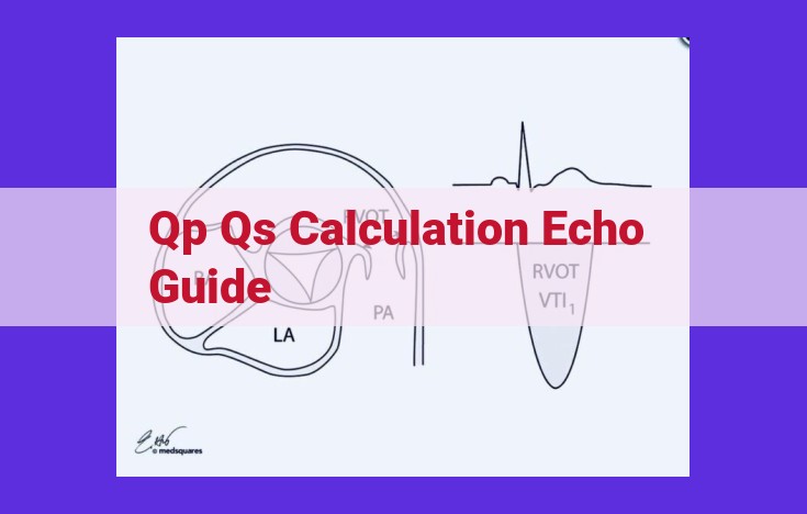
- Continuity Equation Method: A non-invasive technique that utilizes Doppler echocardiography to calculate QP by measuring blood flow velocity and cross-sectional area in the pulmonary artery.
Each method has its strengths and limitations, and the choice of technique depends on the specific clinical situation.
Clinical Applications: Guiding Patient Care
Echocardiography is a valuable tool in diagnosing and managing pulmonary circulation disorders. By accurately assessing QP and QS, physicians can:
- Diagnose pulmonary embolism: Obstruction of the pulmonary artery by a blood clot.
- Evaluate heart failure: A condition where the heart cannot pump effectively, leading to impaired pulmonary circulation.
- Assess lung disease: Conditions such as chronic obstructive pulmonary disease (COPD) and pulmonary fibrosis can affect pulmonary circulation.
Echocardiography provides essential insights into pulmonary circulation, guiding patient care and optimizing treatment outcomes. Understanding the related concepts, such as quadratic index and pressure-volume curve, enhances the accuracy of result interpretation. By leveraging these techniques, physicians can effectively diagnose and manage pulmonary circulation disorders, improving patient health and well-being.
Clinical Applications of Echo in Pulmonary Circulation
Echo’s Diagnostic and Therapeutic Role
Echocardiography provides invaluable insights into pulmonary circulation, facilitating the diagnosis and management of various cardiopulmonary disorders. Its utility extends beyond merely quantifying QP and QS. It can identify and monitor conditions that impair pulmonary blood flow, such as pulmonary embolism, heart failure, and lung disease.
Pulmonary Embolism Detection
In cases of suspected pulmonary embolism, echocardiography plays a crucial role. It can detect right ventricular strain, a telltale sign of elevated pulmonary artery pressure caused by obstructed blood flow. This information aids in prompt diagnosis and timely intervention to prevent life-threatening complications.
Heart Failure Management
Echocardiography is an essential tool in managing heart failure patients. It assesses the structural and functional changes in the right ventricle, which is responsible for pumping blood into the pulmonary circulation. By monitoring right ventricular function, clinicians can tailor treatment plans to alleviate symptoms and improve overall cardiac performance.
Lung Disease Assessment
Echocardiography also aids in the diagnosis and assessment of lung diseases, such as chronic obstructive pulmonary disease (COPD) and pulmonary fibrosis. It evaluates changes in lung tissue, including thickening and scarring, which can significantly impact pulmonary blood flow and gas exchange. This information supports informed decision-making regarding appropriate treatment strategies and patient management.