Throughout history, microscopy, genetics, and molecular biology have provided evidence that mitosis is a continuous process. Microscopy reveals visual cues indicating different stages, while genetic analysis tracks chromosome behavior. Molecular techniques highlight proteins involved in mitosis. This continuous observation has unveiled the complexity of mitosis, its significance in cell biology and health, and the importance of evidence in advancing scientific understanding.
The Continuous Nature of Mitosis: Unraveling the Cellular Symphony
In the realm of cell biology, the dance of mitosis stands out as a remarkable symphony of life, an uninterrupted flow of events that perpetuates the fabric of our existence. Unlike other cellular processes that occur in distinct stages, mitosis unfolds as a continuous stream, a seamless cycle of chromosomal duplication and cellular division.
Mitosis: A Process of Perpetual Motion
At the heart of mitosis lies the concept of continuity. It is a process that transcends the notion of discrete phases, instead flowing like a river, an unstoppable force that ensures the perpetuation of life. Unlike other cellular processes that start and stop, mitosis is a continuous movement, a symphony of events without pauses or interruptions.
The Poetic Language of Mitosis
To capture the essence of mitosis, we turn to metaphors that echo its ceaseless flow. It is a process, a dance, a stream, a cycle. Each of these terms evokes the fluidity and interconnectedness of mitosis, highlighting its uninterrupted nature.
- Process: A sequence of actions or events that follow a natural order, like the harmonious steps of a waltz.
- Flow: A steady movement, like the graceful flow of water in a river, carrying chromosomes through mitosis.
- Stream: A continuous body of flowing water, symbolizing the uninterrupted nature of mitotic events.
- Cycle: A regularly repeating sequence of events, like the cyclical journey of chromosomes as they duplicate, segregate, and re-form.
Embracing Continuity: A New Perspective on Mitosis
By embracing the concept of continuity, we unlock a deeper understanding of mitosis. It is no longer a series of isolated events, but rather a fluid and unified process that unfolds before our eyes. As we delve into the microscope and explore the genetic and molecular evidence, we will discover the intricate details of this cellular masterpiece.
Evidence in the Microscope: Unveiling the Secrets of Mitosis
Microscopy has played a pivotal role in unraveling the intricate details of mitosis. Through the lens, researchers have witnessed the continuous flow of this remarkable process, capturing visual cues that reveal the distinct stages of mitosis.
Microscopy techniques, such as phase-contrast and fluorescence microscopy, allow scientists to observe living cells in real-time, providing a glimpse into the dynamic nature of mitosis. As the cell prepares to divide, microtubules, the cellular filaments responsible for chromosome segregation, become visible within the cytoplasm. These microtubules form a spindle-shaped structure that guides and separates the duplicated chromosomes.
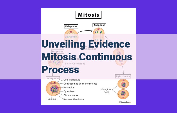
During interphase, the stage preceding mitosis, chromosomes are duplicated, resulting in two sister chromatids held together by a central structure called the centromere. As mitosis progresses, prophase marks the condensation and shortening of chromosomes, making them more visible under the microscope. The nuclear envelope, which surrounds the nucleus, begins to disintegrate.
Metaphase is characterized by the alignment of chromosomes on the metaphase plate, an imaginary plane that forms in the center of the cell. Each chromosome is attached to microtubules from opposite poles of the spindle apparatus.
In anaphase, sister chromatids separate and migrate toward opposite poles of the cell, guided by the shortening microtubules. Telophase, the final stage of mitosis, is marked by the arrival of chromosomes at the poles, where they decondense and become less visible. New nuclear envelopes form around each set of chromosomes, and cytokinesis, the division of the cytoplasm, follows, resulting in two distinct daughter cells.
Microscopy has not only provided visual evidence of mitosis but has also led to the development of microinjection techniques. By injecting fluorescent dyes that bind to specific cellular components, scientists can visualize the dynamics of mitosis at the molecular level. These techniques have helped uncover the complex interplay of proteins that regulate various aspects of mitosis, including spindle formation, chromosome condensation, and cytokinesis.
The continuous observation of mitosis through microscopy has been a cornerstone in the study of cell division. By witnessing the uninterrupted flow of this process, researchers have gained invaluable insights into the molecular mechanisms that govern the faithful transmission of genetic material.
The Role of Chromosomes in Mitosis: Unraveling the Genetic Evidence
In the realm of mitosis, the meticulous partitioning of genetic material plays a pivotal role. Genetic analysis, with its array of sophisticated techniques, has illuminated the intricate dance of chromosomes during this critical cell division process.
Karyotyping, a technique that captures the chromosomal landscape of a cell, has provided invaluable insights. By arranging chromosomes according to their size and banding patterns, researchers can identify abnormalities that may disrupt mitotic progression. For instance, in conditions like Down syndrome, an extra copy of chromosome 21 disrupts the normal chromosomal complement, leading to developmental disabilities.
Fluorescent in situ hybridization (FISH), another powerful technique, allows researchers to visualize specific chromosomes or gene loci within the cellular milieu. By using fluorescent probes that bind to targeted DNA sequences, FISH enables the tracking of chromosomes throughout mitosis. This technique has revealed intricate details of chromosome condensation, alignment, and separation during mitosis.
By harnessing genetic evidence, scientists have unravelled the essential contribution of chromosomes to mitosis. These elegant techniques have not only enriched our understanding of the continuous nature of mitosis but also highlighted the significance of genetic stability for normal cell function and overall health.
Molecular Evidence: Unraveling the Molecular Machinery of Mitosis
Molecular biology techniques have revolutionized our understanding of mitosis, providing profound insights into the intricate molecular mechanisms that orchestrate this essential cellular process. Advanced technologies, such as DNA sequencing and protein analysis, have allowed researchers to decode the genetic blueprint and identify key proteins crucial for spindle formation, chromosome condensation, and cytokinesis.
DNA sequencing revealed the genetic sequences that encode proteins involved in spindle formation. These proteins act as the structural scaffold, guiding the precise segregation of chromosomes during metaphase. Protein analysis identified molecular motors like kinesins and dyneins that power chromosome movement along the mitotic spindle.
Furthermore, the identification of cyclins and cyclin-dependent kinases (CDKs) shed light on the temporal regulation of mitosis. These proteins form complexes that govern the transitions between different mitotic phases. Cyclins accumulate and activate CDKs at specific stages, ensuring the orderly progression of mitosis.
Molecular evidence also unraveled the molecular basis of chromosome condensation. Proteins like condensins and cohesins play vital roles in compacting chromosomes during prophase. By stabilizing the condensed state, they prevent premature chromosome segregation.
Additionally, understanding the molecular mechanisms of cytokinesis, the final stage of mitosis, has been enhanced by molecular biology techniques. The identification of proteins like myosin and actin revealed the cellular machinery responsible for cleaving the cell into two daughter cells.
These molecular discoveries have provided a detailed understanding of the intricate processes that drive mitosis. By dissecting the molecular mechanisms, researchers have gained valuable insights into the regulation, control, and potential therapeutic targets for mitosis-related diseases. The continuous pursuit of molecular evidence continues to unveil the complexity and significance of mitosis.
Unveiling the Complexity of Mitosis: A Journey of Continuous Observation and Discovery
As scientists peered through microscopes, they witnessed the mesmerizing dance of cells undergoing mitosis, a continuous and intricate process. With each observation and piece of evidence gathered, the tapestry of this cellular marvel unraveled.
The Challenges of Unraveling Mitosis
Deciphering the intricacies of mitosis posed significant challenges. Its rapid pace and the elusive nature of cellular structures required researchers to develop innovative techniques. Microscopy became a crucial tool, allowing scientists to capture fleeting moments and study living cells in real-time.
Advancements and Breakthroughs
Technological advancements revolutionized the study of mitosis. Karyotyping, fluorescence in situ hybridization (FISH), and other genetic methods illuminated chromosome behavior. Molecular biology techniques unveiled the molecular machinery orchestrating this complex process. Scientists discovered key proteins responsible for spindle formation, chromosome condensation, and cytokinesis.
Unraveling the Tapestry of Mitosis
The cumulative evidence woven together a comprehensive narrative of mitosis. Researchers recognized its continuous nature, a seamless flow devoid of distinct stages. The microscope served as a window into this dance, revealing visual cues indicative of the different phases of mitosis.
The Importance of Ongoing Research
Despite the remarkable progress, the journey to unravel mitosis is far from over. Challenges remain, such as understanding the precise timing and coordination of events. Ongoing research promises to further deepen our comprehension of this cellular symphony.
Mitosis, a fundamental process in cell biology, holds profound implications for human health and disease. Understanding its intricacies is essential for deciphering the mechanisms behind genetic disorders, cancer, and developmental abnormalities. The continuous observation and accumulation of evidence have been the driving forces behind our ever-evolving understanding of this cellular masterpiece.