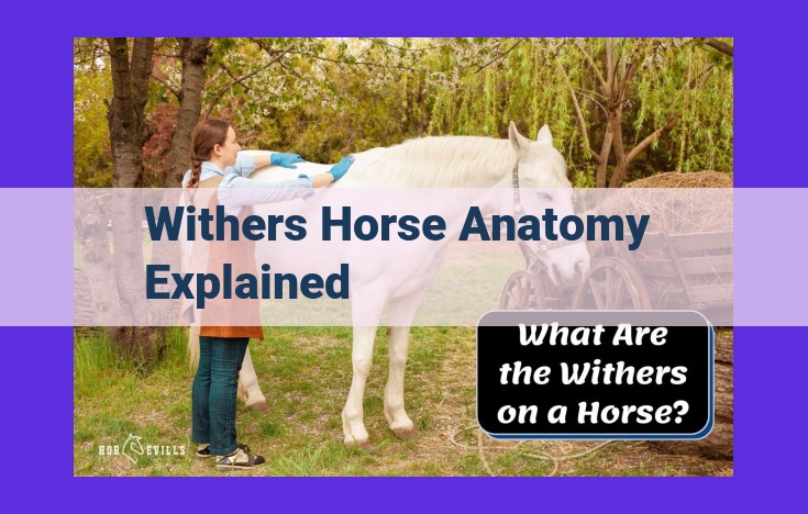
The withers, the highest point of a horse’s back at the base of the neck, are formed by the spinous processes of specific vertebrae. These vertebrae connect to the scapula, which interacts with the withers during movement. The humerus, radius, and ulna are essential for shoulder and forearm movement. Muscles such as the trapezius, rhomboideus, supraspinatus, and infraspinatus play vital roles in shoulder movement and scapula stabilization. Understanding the withers’ anatomy is crucial for assessing and managing various musculoskeletal conditions affecting horses.
Understanding the Withers: The Keystone of Equine Anatomy
In the graceful architecture of a horse, the withers stand as a prominent landmark, a pivotal point at the confluence of the neck and back. These prominent elevations are not merely an aesthetic endowment but play a crucial role in the structural integrity and movement of this majestic creature.
At the very core of the withers lies a complex tapestry of vertebrae, specifically the spinous processes of the dorsal thoracic vertebrae. These vertebrae, stacking one upon another like ancient building blocks, protrude dorsally to form the framework upon which the withers perch. Like the keystone in an arch, these spinous processes anchor the withers, providing the stability and support required for the horse’s every stride.
The withers are not an isolated entity but an integral part of a larger musculoskeletal system. They form the transition zone between the axial skeleton (backbone) and the appendicular skeleton (limbs). The scapula, a triangular blade-like bone, nestles against the withers and provides a pivotal connection to the forelimbs. This connection is essential for transmitting the force generated by the hindquarters to the forelegs, allowing the horse to propel itself forward with elegance and efficiency.
Vertebrae: The Foundation of the Withers
The withers, the prominent ridge at the base of a horse’s neck, are a crucial part of its anatomy. They provide structural support and serve as an attachment point for essential muscles involved in movement. At the heart of the withers lie specific vertebrae, whose spinous processes play a pivotal role in its formation.
The spinous processes extend backward from the vertebrae, forming the dorsal aspect of the withers. These processes are particularly prominent in the thoracic vertebrae, specifically the seventh cervical and first four thoracic vertebrae. The height and angle of the withers are determined by the length and inclination of these spinous processes.
Interlocking facets on the spinous processes provide stability and strength to the withers. They prevent excessive movement and ensure the withers’ structural integrity. Additionally, the intervertebral discs between the vertebrae provide cushioning and flexibility, allowing for controlled movement during activities such as jumping or galloping.
The vertebrae and their spinous processes work in concert to create a solid foundation for the withers, which in turn provides a stable base for the attachment of muscles and the support of the horse’s weight during movement.
The Scapula and Its Connection to the Withers
The scapula, also known as the shoulder blade, is an integral component of the equine musculoskeletal system. It connects the withers, the highest point of the horse’s back, to the axial skeleton and plays a crucial role in shoulder movement.
The scapula is a large, flat bone that lies parallel to the withers. It is attached to the axial skeleton through ligaments and muscles and connects to the humerus (upper arm bone) at the shoulder joint. The glenoid cavity on the scapula forms the socket for the head of the humerus, allowing for a wide range of motion in the shoulder.
During movement, the scapula rotates and glides on the surface of the withers, facilitating the horse’s ability to raise and lower its head and neck. The withers, in turn, provide a stable base for the scapula, preventing excessive movement that could compromise shoulder joint function.
The muscles attached to the scapula, such as the trapezius and rhomboideus, play a key role in stabilizing the scapula and controlling its movement during locomotion. These muscles work together to move the scapula forward and backward, upward and downward, and rotate it to facilitate proper shoulder function.
The Humerus: A Key Player in Shoulder Movement
The humerus is the longest bone in the forelimb of a horse, and it plays a crucial role in both shoulder and elbow movement. It connects the scapula (shoulder blade) to the radius and ulna (forearm bones), forming the shoulder joint.
The humerus has three distinct sections: the proximal (upper) end, the diaphysis (shaft), and the distal (lower) end. The proximal end of the humerus forms the glenoid cavity, which articulates with the glenoid cavity of the scapula to create the shoulder joint. This joint allows for a wide range of motion, including flexion, extension, and rotation.
The diaphysis of the humerus is relatively straight and cylindrical. It provides attachment points for several muscles that control shoulder movement, including the deltoid, biceps, and triceps muscles. The deltoid muscle abducts (lifts away from the body) the shoulder, while the biceps and triceps muscles flex and extend the elbow, respectively.
The distal end of the humerus is wider than the proximal end and has two condyles (rounded projections) that articulate with the radius and ulna. These condyles allow for a hinge-like motion at the elbow joint, enabling the horse to bend and straighten its foreleg.
The humerus is a crucial bone for shoulder and elbow movement in horses. Its unique structure and connections to surrounding muscles allow for a wide range of motions that are essential for locomotion, athletic performance, and overall well-being.
Radius and Ulna: Function in Forearm Movement:
- Describe the location and connection of the radius and ulna in the forearm.
- Explain their role in wrist and elbow movement.
Radius and Ulna: The Dynamic Duo of Forearm Movement
The radius and ulna, two long bones that connect the humerus (upper arm bone) to the wrist, play a crucial role in the incredible mobility of your horse’s forelimbs.
Location and Connection
Imagine the radius and ulna as a parallel pair, situated side-by-side in your horse’s forearm. The radius lies lateral (on the outer side), while the ulna occupies the medial (inner) position. Together, they form a hinge-like joint at the elbow and a pivot joint at the wrist.
Role in Forearm Movement
The radius and ulna are responsible for the following movements:
-
Elbow: The radius and ulna extend and flex (bend) the elbow joint, allowing your horse to lift and lower its foreleg.
-
Forearm Rotation: These bones work together to pronate and supinate the forearm. Pronation involves turning the palm downward, while supination rotates the palm upward.
-
Wrist: The radius and ulna articulate with the carpal bones (wrist bones) to facilitate flexion and extension of the wrist joint. This enables your horse to flex its fetlock and cannon bone.
The radius and ulna, working in harmony, provide your horse with the precision and flexibility it needs in its forelimbs. These bones allow for a wide range of movements, from gentle strides to explosive jumps. By understanding their vital functions, you gain a deeper appreciation for the intricate mechanics behind your horse’s graceful movements.
The Trapezius Muscle and Its Role in Shoulder Movement
In the equestrian world, understanding the intricate muscular anatomy of horses is crucial for optimizing performance and preventing injuries. One of the most important muscles involved in shoulder movement is the trapezius muscle. This robust muscle plays a vital role in moving the shoulder and stabilizing the scapula, ensuring smooth and coordinated motion.
Origin and Insertion Points
The trapezius muscle, originating from the midline of the neck’s dorsal surface, extends down the entire length of the withers and inserts on the lateral aspect of the spine of the scapula, connecting the axial skeleton to the shoulder. Due to its wide origin, the trapezius muscle can exert significant force on the scapula and shoulder joint.
Functions of the Trapezius Muscle
The trapezius muscle performs a variety of functions related to shoulder movement:
-
Elevation of the scapula: By contracting, the trapezius muscle lifts the scapula upward, creating space for the humerus to move freely within the shoulder joint. This action is crucial for proper shoulder flexion and extension.
-
Adduction of the scapula: The trapezius muscle also plays a role in pulling the scapula towards the midline of the body, bringing the shoulder joint closer to the body’s center of gravity. This movement stabilizes the scapula during weight-bearing activities, such as walking or running.
-
Rotation of the scapula: Additionally, the trapezius muscle assists in rotating the scapula, allowing for greater range of motion in the shoulder joint. This rotation is essential for proper function of the supraspinatus and infraspinatus muscles, which are involved in shoulder abduction and external rotation, respectively.
Stabilization of the Scapula
Beyond its role in shoulder movement, the trapezius muscle also serves as a stabilizer for the scapula. By maintaining tension across the scapula, it helps to prevent excessive movement during locomotion and other activities. This stabilization ensures that the scapula remains in a stable position relative to the thorax, allowing for optimal muscle function and joint mechanics.
By understanding the anatomy and functions of the trapezius muscle, horse owners and riders can gain a deeper appreciation for the complexity of equine shoulder mechanics. This knowledge can empower them to make informed decisions regarding training, conditioning, and injury prevention, ultimately contributing to the well-being and athletic performance of their horses.
The Rhomboideus Muscle: A Key Player in Shoulder Stability
Tucked beneath the trapezius muscle, the rhomboideus muscle, like an unsung hero, plays a crucial role in orchestrating the smooth and stable movement of your shoulder. Its fibers gracefully arch from the vertebrae of your spine to the scapula, the triangular bone that forms the foundation of your shoulder joint.
With each contraction, the rhomboideus muscle retracts (pulls back) the scapula, stabilizing it against the rib cage. This unwavering support provides a solid base for the shoulder joint to glide and rotate effortlessly. Without the rhomboideus’s steady hand, your scapula would float aimlessly, compromising the efficiency and precision of your shoulder movements.
Origin and Insertion: Originating from the spinous processes of the thoracic vertebrae T2 to T5, the rhomboideus muscle’s fibers elegantly converge to form two distinct bundles. The rhomboideus major, the larger and more prominent of the duo, inserts onto the medial border of the scapula, while the rhomboideus minor, its smaller counterpart, attaches to the scapula’s vertebral border.
Function: The rhomboideus muscle is primarily responsible for retracting the scapula, effectively pulling it towards the spine. This movement is essential for stabilizing the shoulder joint and facilitating a wide range of arm movements. During shoulder elevation, for instance, the rhomboideus muscle works in harmony with other muscles to lift the scapula upwards, creating the necessary space for the arm to swing freely overhead.
Furthermore, the rhomboideus muscle plays a pivotal role in adducting (drawing towards the midline) the scapula. This action is particularly important in movements that require the arms to be brought towards the body, such as reaching forward or hugging oneself. By adducting the scapula, the rhomboideus muscle helps to stabilize the shoulder joint and prevent excessive outward rotation.
Strengthening the Rhomboideus Muscle: Regular exercises that target the rhomboideus muscle can enhance its strength and stability, leading to improved shoulder health and performance. Exercises like shoulder retractions and dumbbell rows are excellent ways to engage this muscle and build its resilience.
The Supraspinatus Muscle: A Key Player in Shoulder Abduction
In the intricate symphony of muscles that control our shoulder movements, the supraspinatus muscle plays a pivotal role. Its job? To lift our arms away from our bodies, allowing us to perform a wide range of everyday activities.
Located in the shoulder joint, the supraspinatus muscle originates from the suprascapular fossa on the scapula (shoulder blade). It then inserts onto the greater tubercle of the humerus (upper arm bone).
When the supraspinatus muscle contracts, it abducts the arm, lifting it away from the body. This action is essential for activities like reaching overhead to grab something or brushing our hair.
The supraspinatus muscle works in conjunction with other shoulder muscles to achieve full range of motion. For instance, the deltoid muscle assists in abduction, while the rotator cuff muscles stabilize the shoulder joint.
Understanding the supraspinatus muscle’s role is not only important for overall shoulder function but also for preventing injuries. Overuse or improper use of the supraspinatus muscle can lead to tendinitis or tears, which can cause pain and limit mobility.
By engaging in regular stretching and strengthening exercises, we can help keep our supraspinatus muscles healthy and strong, ensuring that our shoulders remain flexible and ready for action.
The Infraspinatus Muscle: A Key Player in External Shoulder Rotation
The infraspinatus muscle is an integral part of the shoulder complex, playing a crucial role in external rotation – the movement of the arm away from the body. Understanding its anatomy and function is essential for maintaining optimal shoulder health.
Located deeply within the shoulder joint, the infraspinatus originates from the dorsal surface of the scapula. It is attached to the spine and lateral border of the shoulder blade. From there, the infraspinatus muscle runs laterally across the back of the shoulder, inserting onto the greater tubercle of the humerus.
This muscle’s primary function is to externally rotate the shoulder joint. When it contracts, it pulls the humerus outward, rotating the arm away from the midline of the body. External rotation is essential for everyday activities such as reaching overhead, pouring liquids, and throwing objects.
In addition to its role in external rotation, the infraspinatus muscle also contributes to stabilizing the shoulder joint. It works with other rotator cuff muscles to keep the humeral head centered within the glenoid cavity of the scapula. This stability is crucial for preventing dislocations and ensuring proper movement of the shoulder.
Therefore, maintaining a healthy infraspinatus muscle is essential for optimal shoulder function. Regular exercise and proper body mechanics can help strengthen this muscle and reduce the risk of shoulder injuries. If you experience pain or discomfort in your shoulder, it is important to consult a healthcare professional to rule out any underlying issues and receive appropriate treatment.